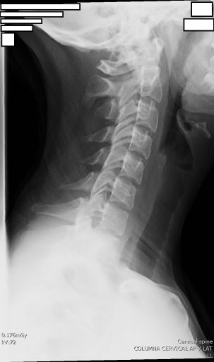
LAT CERVICAL
Lateral Projection of Cervical Spine • Alignment and Structure Evaluation
Exposure Factors
Increased distance: Greater distance to reduce magnification in lateral projection
Increased Focus-Plate Distance
Purpose: Reduce magnification and improve definition in lateral projection
Comparison: Greater than AP cervical (105-115 cm) to compensate for object-plate increase
Visible Anatomical Structures
Should be clearly observed:
- Vertebral bodies from C1 to C7
- Spinous processes
- Uncinate processes
- Transverse processes
- Intervertebral disc space
- Pedicles
- Laminae
- Intervertebral joints
- Skull base
Plate Size and Orientation
Longitudinal orientation to cover entire cervical spine from skull base to D1
Patient Positioning
Central Ray Point
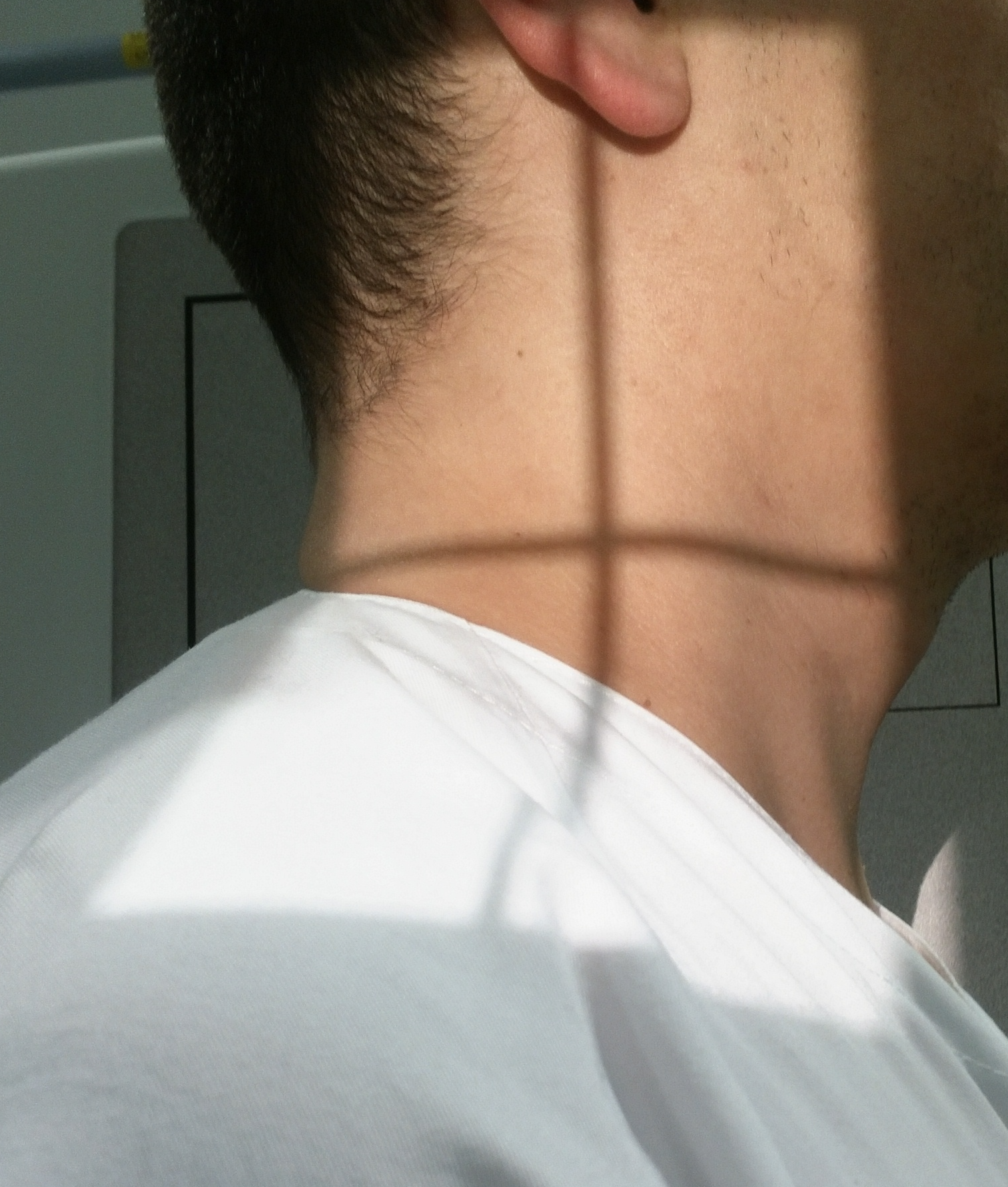
Location: Directed to the fourth cervical vertebra (C4)
Angulation: Horizontal (no angulation)
Direction: Perpendicular to plate and long axis of neck
Optimal Image Characteristics
Vertebrae C1-C7
All visible without overlap
Disc Spaces
Intervertebral spaces open
Overlaps
Jaw separated from vertebrae
Skull Base
Clearly visible
Processes
Spinous and transverse defined
Alignment
Physiological cervical curvature
BREATHING INSTRUCTIONS
"At the end of expiration, suspend breathing"
- Perform exposure at end of expiration
- Shoulders in lowest possible position
- Allows better visualization of C7-T1
- Reduces movement during exposure
Technique: Patient exhales completely and holds breath
Common Technical Challenges
Frequent problems in cervical lateral projection:
- Jaw-vertebrae overlap due to insufficient chin elevation
- Shoulders not lowered hiding C6-C7-T1 vertebrae
- Head/trunk rotation creating oblique images
- Excessive magnification due to insufficient focus-plate distance
- Poor C7-T1 visualization due to wide shoulders or muscular patients
Solution: Actively lower shoulders and use increased focus-plate distance (120-150 cm)
Patient in Supine Position (Stretcher)
Procedure for patients who cannot mobilize:
- Place chassis vertically longitudinally beside the neck
- Center chassis at C4
- Immobilize chassis with sandbags on sides
- Alternative: secure with adhesive tape
- Horizontal central ray directed to C4
Trauma priority: Do not move the patient, adapt technique to their position
Patient Instructions
"At the end of expiration, suspend your breathing"
Complete sequence:
1. Place shoulder against bucky
2. Lower shoulders maximally
3. Slightly elevate chin
4. Exhale completely
5. Hold breath
6. Maintain position without moving